Digital Dentistry: Intraoral Scanning and Computer-Designed Restorations
This page explains digital dentistry: what an intraoral scan replaces, research on its comfort and accuracy, how a crown can be designed on screen, and where conventional impressions still do better.
5.0 from 550+ Google reviews
Open 8am to 8pm, seven days
Free parking at the door
CQC & GDC registered
No referral needed
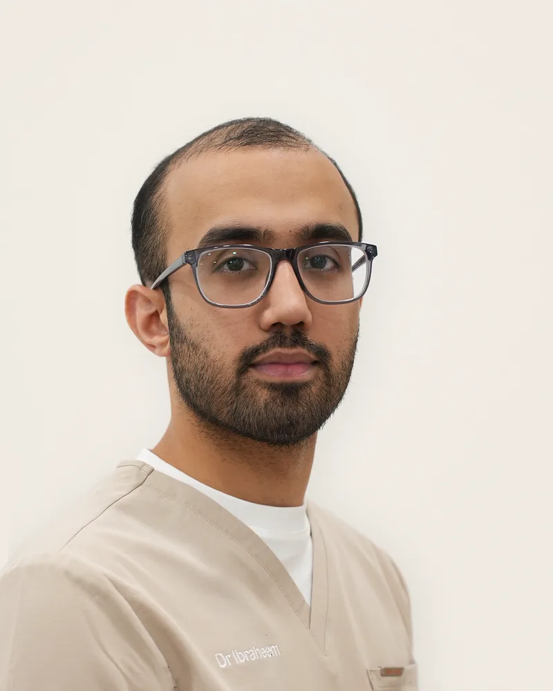
Clinically reviewed by Dr Ibraheem Ijaz, dentist · GDC 301711
Medical disclaimer: This page is for general information only. It does not replace an examination or treatment by a dentist.
Dr Ibraheem Ijaz, the practice's principal dentist and a General Dental Practitioner (GDC 301711), reviewed this page. His qualifications are BDS, BSc (Hons), MSc (Leeds), PG Dip Implant Dentistry and PG Cert Restorative and Aesthetic Dentistry.
What is digital dentistry?
Digital dentistry is the recording, planning and making of dental treatment on a computer, usually starting from a 3D scan of the teeth.
An intraoral scanner is a handheld wand that projects light onto the teeth and gums. A 2017 review in BMC Oral Health (Mangano and colleagues) describes software turning the images into a 3D model, saved as a file such as an STL file. Computer-aided design and manufacture (CAD/CAM) then turn that model into a restoration such as a crown, drawn on screen and milled by machine.
A scan records shape; choosing treatment still rests on a clinical examination.
What does a digital scan replace?
An intraoral scan replaces a conventional impression, a tray of soft, putty-like material held in the mouth until it sets.
A 2020 review (Kihara and colleagues) names the materials as alginate and rubber-like elastomers. Mangano's 2017 review adds that the impression is then cast in plaster to make a model, a step a scan skips.
Are digital impressions comfortable and accurate?
Research suggests patients generally prefer scans, and that scans match conventional impressions in accuracy for crowns and short bridges.
Comfort
A 2018 systematic review (Gallardo and colleagues) found only five eligible studies. Four found the digital technique more comfortable; one found no difference. The authors found clinical studies of patient outcomes lacking.
A 2021 systematic review (Siqueira and colleagues) found scanning generally preferred, whether part or all of an arch was scanned: 10 of the 11 studies that asked patients favoured it, and one found no difference in comfort. The authors noted differing study designs and that patients could not be blinded to the method.
Accuracy
A 2018 systematic review (Ahlholm and colleagues) found digital impressions as accurate as conventional ones for crowns and short bridges. For full-arch impressions, conventional methods were more accurate, and the authors still recommended them there.
A 2022 meta-analysis of studies in patients (Kong and colleagues) found full-arch scans and alginate impressions similarly true, a 3D difference of 0.09 mm, and said scans can be considered a viable alternative to alginate for people with all their natural teeth. It called for more research on other materials.
How are restorations designed digitally?
In a digital workflow, a crown can be drawn on screen from the scan and then milled from a block of ceramic or resin-based material.
In Mangano's 2017 review, clinics equipped for chairside work mill the design themselves; otherwise the scan files go to a dental laboratory.
How do computer-designed restorations perform?
A 2018 systematic review of 2,916 single-tooth ceramic CAD/CAM restorations followed for an average of 7 years (Alves de Carvalho and colleagues) estimated 5-year survival at 89.7%, a failure rate of 2.17% a year.
A 2020 meta-analysis of resin-based (hybrid polymer) and ceramic CAD/CAM crowns (Al-Haj Husain and colleagues) found survival of 99% at up to 2 years and 95% at 3 years or more. Overall success fell from 88% to 77%, a change it did not find significant. Technical success was significantly lower for partial crowns, 64% (95% confidence interval 34% to 86%), than for full crowns, 93%. The authors suggested chairside milling without a technician checking material thickness could have contributed to the partial crowns' complications.
A 2022 review of six studies of zirconia and lithium disilicate crowns made from intraoral scans (Benli and colleagues) found chipping in 1.4% of zirconia and 5% of lithium disilicate crowns, with appearance the most frequent complication for zirconia.
To ask about restorative treatment, book online or call 0114 288 2121.
Where does digital dentistry have the advantage?
Digital methods tend to have the advantage in speed and in sharing the record with the laboratory and the patient.
Ahlholm's 2018 review found scanning faster. Siqueira's 2021 review, within its stated limitations, found scanning faster overall; of the 14 studies that timed it, 11 favoured scanning and three conventional impressions. Gallardo's 2018 review was split on time across five studies, two favouring conventional impressions. Mangano's 2017 review adds a scan checked on screen as it is taken, files sent electronically to the laboratory, and better communication with the patient. Siqueira adds that conventional impressions need disinfecting, which can distort the material.
Where do conventional methods still have the advantage?
Reviews suggest scanners have limits with long spans, edges prepared deep under the gum, and bleeding. For long-span restorations, Mangano's 2017 review said conventional impressions are still indicated.
Mangano's review also said deep margin lines, or bleeding, can be hard to capture, though it added that with careful technique a good scan is still possible even in difficult cases. Siqueira's 2021 review cites a trial in which margins prepared deep under the gum gave less accurate scans, and notes that errors can build up across a full-arch scan from saliva, shiny restorations and movable gum tissue. Kihara's 2020 review considered cross-arch bridges difficult to make from scans, and Mangano adds a learning curve and the cost of a scanner.
How is digital technology used across treatments?
Intraoral scans are used in restorative, cosmetic, orthodontic and implant dentistry.
In restorative dentistry, Mangano's 2017 review judged scanners accurate enough for inlays, onlays, single crowns and short bridges; the steps of having a crown fitted have their own page.
In cosmetic dentistry, Mangano's review notes scans can be used for digital smile design, which has its own page on how a planned change is simulated, reviewed and approved before treatment starts. Colour on a scan, the review says, matters mainly for talking with the patient; dental shade matching is covered separately.
In orthodontics, scans are the starting point for clear aligners, and how an aligner scan becomes a treatment plan is explained separately. In implant dentistry, Mangano's 2017 review describes scans overlaid on CBCT (3D X-ray) images for guided surgery, a use it said was only in its infancy; CBCT scans in implant planning have their own page.
What imaging equipment is on site at Deepcar Dental Care?
Imaging equipment on site at Deepcar Dental Care includes:
- Intraoral scanning (scanner made by Shining 3D)
- Digital X-rays
- OPG machine
- Intraoral camera
Where can you read more about restorative dentistry technology?
Root canal equipment is explained on the page about rubber dam, rotary instruments and apex location, one of the options in this section.
Restorative dentistry saves and rebuilds natural teeth rather than replacing them with implants.
Frequently asked questions
Does an intraoral scan use X-rays?
No. An intraoral scanner records the surface of the teeth and gums with light. A 2017 review in BMC Oral Health describes scanners projecting laser or structured light onto the teeth. Imaging that shows roots and bone, such as dental X-rays and CBCT scans, does use radiation.
How long does an intraoral scan take?
A full-arch scan may take 3 to 5 minutes, similar to a conventional impression, according to a 2017 review in BMC Oral Health. It says the main time saving appears to come afterwards, with no plaster model to pour. A 2021 systematic review, within its stated limitations, found scanning faster overall than conventional impressions, including full-arch scans.
Do all intraoral scanners give the same accuracy?
No. A 2017 review in BMC Oral Health found only a few studies comparing scanners, but concluded that different scanners have different accuracy, some seeming suited only to single or short-span restorations. A 2020 review also reported that the brightness and colour temperature of the lighting affected scanner accuracy.
Is a scan easier if you gag during impressions?
It may be. A 2017 review in BMC Oral Health notes that some people with a strong gag reflex appear unable to tolerate conventional impressions. A 2018 systematic review reported less nausea and anxiety with scanning in four of its five studies. If you gag easily, tell your dentist before any impression or scan.
What is an STL file in dentistry?
An STL file is a standard 3D model format. A 2017 review in BMC Oral Health explains that open scanners save files such as STL that other design software and laboratories can read, while closed systems save their own formats and may charge a fee to unlock them.
Book a consultation.
Online in under a minute, or call and speak to a real person.
You can call Deepcar Dental Care or book online. The practice is open 8am to 8pm, Monday to Sunday. Our patient reviews show what patients say.
334 Manchester Road
Deepcar, Sheffield S36 2RH
0114 288 2121
Contact us
Opening hours
Open 8am to 8pm, seven days
Free parking at the door